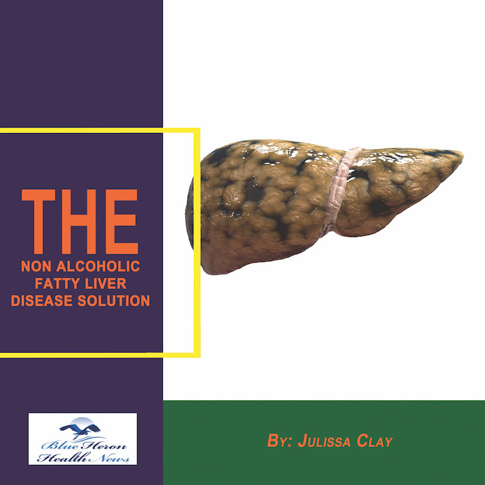
The Non Alcoholic Fatty Liver Strategy™ By Julissa Clay The problem in the fatty liver can cause various types of fatal and serious health problems if not treated as soon as possible like the failure of the liver etc. The risks and damage caused by problems in the non-alcoholic liver with fat can be reversed naturally by the strategy provided in this eBook. This 4-week program will educate you about the ways to start reversing the risks and effects of the disease of fatty liver by detoxing your body naturally. This system covers three elements in its four phases including Detoxification, Exercise, and Diet.
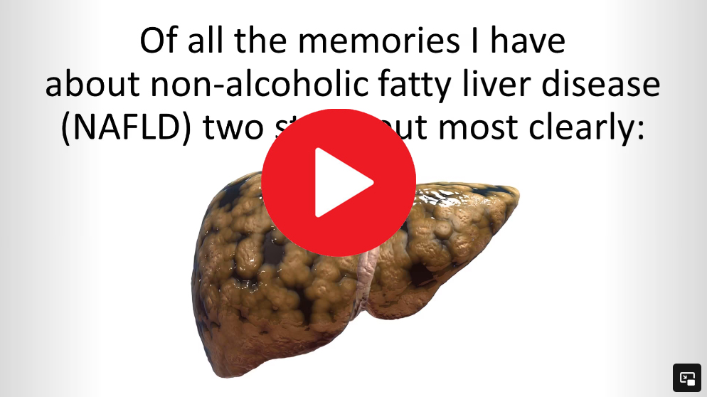
How is Fatty Liver Disease Diagnosed?
Fatty Liver Disease, also known as hepatic steatosis, is diagnosed through a combination of methods that include medical history, physical examinations, blood tests, imaging studies, and sometimes a liver biopsy. Here’s a detailed overview of the diagnostic process:
1. Medical History and Physical Examination:
- Medical History: The doctor will inquire about the patient’s medical history, including alcohol consumption, medications, family history of liver diseases, and risk factors such as obesity, diabetes, or high cholesterol.
- Physical Examination: The doctor may check for signs of liver disease, such as an enlarged liver, jaundice (yellowing of the skin and eyes), or fluid retention in the abdomen.
2. Blood Tests:
- Liver Function Tests (LFTs): These tests measure enzymes and proteins in the blood that indicate liver health. Elevated levels of certain liver enzymes, such as ALT (alanine aminotransferase) and AST (aspartate aminotransferase), can suggest liver inflammation or damage.
- Lipid Profile: To check for high levels of cholesterol and triglycerides, which are often associated with fatty liver disease.
- Glucose Levels: To assess for diabetes or prediabetes, which are risk factors for non-alcoholic fatty liver disease (NAFLD).
3. Imaging Studies:
- Ultrasound: The most common imaging test used to detect fatty liver. It can show the presence of fat in the liver but cannot determine the extent of inflammation or fibrosis.
- Computed Tomography (CT) Scan or Magnetic Resonance Imaging (MRI): These imaging techniques can also detect fat in the liver and provide more detailed images compared to an ultrasound.
- Transient Elastography (FibroScan): A specialized ultrasound technique that measures liver stiffness, which can indicate fibrosis or scarring of the liver.
4. Liver Biopsy:
- This is the most definitive way to diagnose and stage fatty liver disease, particularly in cases where the diagnosis is uncertain or there is a need to assess the extent of liver damage. A small sample of liver tissue is taken and examined under a microscope to detect fat, inflammation, and fibrosis.
5. Non-Invasive Tests:
- FibroTest, FibroMeter, or Enhanced Liver Fibrosis (ELF) Test: These blood tests combine different biomarkers to estimate the level of fibrosis in the liver. They are less invasive alternatives to a liver biopsy but may not be as definitive.
6. Other Assessments:
- The doctor may also evaluate other factors, such as lifestyle, diet, and exercise habits, to determine the presence of metabolic syndrome, which is often associated with fatty liver disease.
Diagnosis of fatty liver disease usually starts with non-invasive methods like blood tests and imaging, but a liver biopsy may be recommended if more detailed information is needed.
Certainly! Here’s a more detailed breakdown of the methods used to diagnose Fatty Liver Disease:
1. Medical History and Physical Examination
- Medical History: The physician will ask about:
- Alcohol Consumption: Heavy alcohol use is a major risk factor for alcoholic fatty liver disease (AFLD). For non-alcoholic fatty liver disease (NAFLD), even moderate drinking could still be relevant.
- Medication Use: Certain medications, such as corticosteroids, tamoxifen, or methotrexate, can contribute to fatty liver.
- Family History: A history of liver disease, obesity, or diabetes in the family may increase the risk of developing fatty liver disease.
- Lifestyle Factors: Diet, exercise habits, and weight history will be discussed. Diets high in saturated fats and sugars, along with sedentary behavior, are common contributors to NAFLD.
- Associated Conditions: The doctor will check for the presence of obesity, type 2 diabetes, hyperlipidemia (high cholesterol and triglycerides), or metabolic syndrome.
- Physical Examination: The physician will look for signs such as:
- Hepatomegaly: Enlargement of the liver, which can be detected by palpation (feeling the liver area) during a physical exam.
- Jaundice: Yellowing of the skin and eyes, indicative of liver dysfunction.
- Spider Angiomas: Small, spider-like blood vessels visible under the skin, which can be a sign of advanced liver disease.
- Ascites: Accumulation of fluid in the abdomen, which can occur in cirrhosis, an advanced stage of liver disease.
- Skin Changes: Darkened skin (acanthosis nigricans), particularly around the neck or armpits, can be a sign of insulin resistance, which is closely associated with NAFLD.
2. Blood Tests
- Liver Function Tests (LFTs):
- Alanine Aminotransferase (ALT): Elevated levels indicate liver cell injury. ALT is more specific to the liver compared to AST.
- Aspartate Aminotransferase (AST): Elevated AST can indicate liver or muscle damage.
- Alkaline Phosphatase (ALP): High levels may suggest bile duct problems or liver damage.
- Gamma-Glutamyl Transferase (GGT): Increased levels are associated with liver disease and excessive alcohol consumption.
- Bilirubin: Elevated levels may indicate liver dysfunction or bile duct issues.
- Albumin and Total Protein: These measures can indicate the liver’s synthetic function, with low levels suggesting advanced liver disease.
- Fasting Blood Glucose and HbA1c: These tests evaluate blood sugar levels and help diagnose diabetes or prediabetes, both of which are risk factors for NAFLD.
- Lipid Profile:
- Triglycerides: High levels are commonly seen in individuals with fatty liver disease.
- Low-Density Lipoprotein (LDL) Cholesterol: Elevated LDL is often found in those with NAFLD.
- High-Density Lipoprotein (HDL) Cholesterol: Low HDL levels are a component of metabolic syndrome, which is linked to NAFLD.
3. Imaging Studies
- Ultrasound:
- Basic Ultrasound: This is often the first imaging test performed. It can detect the presence of fat in the liver (appears as a brighter liver on ultrasound), but it cannot quantify the amount of fat or assess fibrosis.
- Advantages: Non-invasive, widely available, and cost-effective.
- Limitations: Cannot differentiate between simple steatosis (fat accumulation) and more severe forms like steatohepatitis (fat plus inflammation).
- Computed Tomography (CT) Scan:
- Basic CT: Can detect liver fat by showing a lower density of liver tissue compared to other organs. However, CT is not typically used as a first-line tool due to radiation exposure.
- Contrast-Enhanced CT: Sometimes used to assess liver architecture, but not specifically for fatty liver detection.
- Magnetic Resonance Imaging (MRI):
- Proton Density Fat Fraction (PDFF) MRI: A specialized type of MRI that quantifies the percentage of fat in the liver. It’s more accurate than ultrasound but more expensive.
- Magnetic Resonance Elastography (MRE): Measures liver stiffness and can assess both fat content and fibrosis.
- Transient Elastography (FibroScan):
- Principle: Uses a pulse of ultrasound to measure liver stiffness. The speed of the wave correlates with liver stiffness, with higher stiffness indicating more fibrosis.
- Advantages: Non-invasive and can provide an estimate of fibrosis without a biopsy.
- Limitations: May be less accurate in obese individuals.
4. Liver Biopsy
- Procedure:
- How It’s Done: A needle is inserted into the liver, usually under ultrasound guidance, to remove a small tissue sample.
- Microscopic Examination: The tissue is examined for the presence of fat, inflammation, hepatocyte ballooning (swelling of liver cells), and fibrosis (scarring).
- Staging: Biopsy remains the gold standard for staging liver fibrosis and distinguishing between simple steatosis and non-alcoholic steatohepatitis (NASH), a more severe form of NAFLD that can progress to cirrhosis.
- Risks:
- Pain: The most common side effect, though usually mild.
- Bleeding: Rare but can occur at the biopsy site.
- Infection: Very rare, but possible.
- When It’s Used: Typically reserved for cases where:
- The diagnosis is uncertain after non-invasive tests.
- There’s a need to assess the severity of liver disease.
- There’s a suspicion of cirrhosis or when deciding on treatment options.
5. Non-Invasive Tests for Fibrosis
- FibroTest, FibroMeter, and Enhanced Liver Fibrosis (ELF) Test:
- How They Work: These blood tests use combinations of biomarkers to estimate liver fibrosis. They can be used to monitor disease progression and response to treatment.
- Advantages: Non-invasive, safer than a liver biopsy, and can be repeated as needed.
- Limitations: They may not be as accurate as a liver biopsy and can sometimes yield false positives or negatives.
6. Assessment of Associated Conditions
- Metabolic Syndrome:
- Diagnosis Criteria: Includes central obesity, high blood pressure, high fasting glucose, high triglycerides, and low HDL cholesterol.
- Importance: Metabolic syndrome is strongly linked to NAFLD and indicates a higher risk for cardiovascular disease.
- Insulin Resistance and Diabetes:
- Homeostasis Model Assessment of Insulin Resistance (HOMA-IR): A calculation based on fasting insulin and glucose levels to assess insulin resistance.
- Oral Glucose Tolerance Test (OGTT): Assesses how the body handles glucose over time and can diagnose diabetes or prediabetes.
Summary:
The diagnosis of fatty liver disease is typically a stepwise process, starting with a detailed medical history and physical examination, followed by blood tests and imaging studies. Liver biopsy remains the gold standard for definitive diagnosis and staging, especially in cases where more severe liver disease is suspected. Non-invasive tests and imaging are increasingly used to monitor the disease progression and guide treatment without the need for biopsy in all cases.
The Non Alcoholic Fatty Liver Strategy™ By Julissa Clay The problem in the fatty liver can cause various types of fatal and serious health problems if not treated as soon as possible like the failure of the liver etc. The risks and damage caused by problems in the non-alcoholic liver with fat can be reversed naturally by the strategy provided in this eBook. This 4-week program will educate you about the ways to start reversing the risks and effects of the disease of fatty liver by detoxing your body naturally. This system covers three elements in its four phases including Detoxification, Exercise, and Diet.